Tests on our GcMAF
Our Assays
-

- GcMAF Products
- Our Assays
- External Independent Assays
- Aids Case Report No 1
- Dr Jeff Bradstreet in GcMAF's laboratory
Our GcMAF is made in highly professional sterile laboratories by GMP-trained scientists. We use sterile, certified substrates with traceability, and sterile, certified equipment.
Every batch of our human GcMAF is subjected to internal and external assays.
Its has the following production assays:
1. Sterility – to USP and Ph Eur sterility standards performed externally.
2. Endotoxin test to confirm the absence of endotoxins in the sample. Its 0.02 to 0.03, the bottom limit of detection. (10.0 is the max acceptable)
3. Protein Quantification using BCA Protein assay
4. Electrophoresis Silver Stained SDS Page for product identification;
5. Electrophoresis Western blot probed with biotin labelled Helix Pomatia Lectin (binding directly to the terminal N-acetylgalactosaminyl present in Gc MAF).
6. Electrophoresis Western Blot probed with Anti Vitamin D Binding Protein (specific to Gc Globulin and Gc MAF)
7. RAW 264.7 live macrophage cell based proliferation assay for activity, ie potency,
8. Breast carcinoma phagocytosis activity assay. Macrophages are added to live MCF7 breast cancer cells; nothing happens. We add our GcMAF; withIn 72 hours the macrophages are observed to phagocytise (eat and destroy) the cancer cells.
9. Third activity assay: we add our GcMAF to MCF7 cancer cells without macrophages. On addition of GcMAF a cell morphology change is observed In 72 hours where cancer cells adopt a normal cell morphology. (Experiment first performed by Professor Ruggiero’s team (with our GcMAF) and published January 2012)
The molecules of GcMAF we prepare are identical to those made by the human body.
We have carried out about 330 assays on our GcMAF. Our GcMAF is by far the most heavily tested in the history of GcMAF. Most others have zero tests or one test, and no others have activity tests.
External independent assays our GMAF has passed in Universities/laboratories include
USA – Activity: Macrophage J774 murine cell line activity assay - Ohio University (Feb 2010 onwards)
In Europe – Activity: Chorioallantoic Membrane (CAM) Assay (First 16.05.2010
In Europe – Activity: Destruction of cultured human breast cancer cells
In Europe – 17 day sterility assay: 7 days at 30 degrees C, 7 days at 25 deg, + sub culture aerobic, anaerobic, at 25 degrees. All results negative, “clear as a bell” HPA (First 18.01.2011, monthly)
Our GcMAF has twice been shown by independent laboratories to be as potent as samples provided by Dr Yamamoto. But we have have far better testing.
Activity assays are vital - GcMAF must be proven to exist, be sterile, and most importantly, be active, and activity assays can only be done with living cells. If you have any questions about assays, email or call us by clicking on “Contact” at the top.
Independent longevity assays of our GcMAF:
Test after 8 months stored in a freezer: Our GcMAF had negligible reduction in activity. 2nd Feb 2011
CAM assay: Kept at room temperature for 10 days: lost 15 percent of its activity. May 2010
“Badly kept” assay: 7 degrees in frequently opened fridge, shots removed by needle: Good activity after 4 weeks, nearly zero activity after 8 weeks. 11th Feb 2011
“Well kept assay” at +4 degrees in fridge: reduced, but still good activity at 8 weeks. 11th Feb 2011
Research papers:
Ours is the publicly available only GcMAF that has been used in research papers (eleven of them) for example:
“Gc protein-derived macrophage-activating factor (GcMAF) stimulates cAMP formation in human mononuclear cells and inhibits angiogenesis in chick embryo chorionallantoic membrane assay” published in
“Cancer Immunology and Immunotherapy” in which they kindly used our GcMAF. See the Materials and Methods section. In table 5 they show Yamamoto’s GcMAF at an activity of 17.4, and ours at 18.3, a difference of 5.2 percent, ie almost the same potency.
Cancer Immunol Immunother DOI 10.1007/s00262-010-0953-7
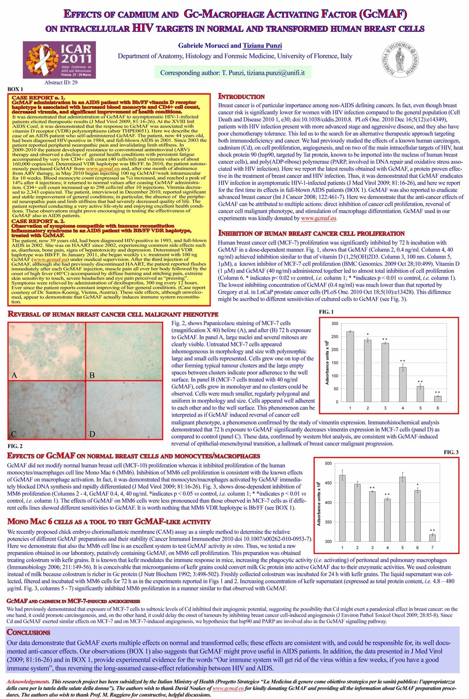
The aids Case Report No 1 is particularly interesting, as is our GcMAF’s ability, in the absence of macrophages, to turn cancer cells back into normal cells (ABCD picture Fig 2.)